


For most of human history, the inner workings of the human body remained a secret. Over the last few centuries, the invention of the microscope and other scientific techniques allowed scientists to learn about the structure and function of our anatomy. Just in the past decade, researchers perfected the art of imaging microstructures while highlighting different cell types with fluorescent colors. In some cases, it helps make it possible to see the glow with the naked eye.
That’s what happened when Kjeld Møllgård, an 80-year old neuroscientist, was dissecting mice to study the structure of microscopic tunnels surrounding the brain—and spotted something unexpected.
This was part of a research study led by Maiken Nedergaard, a professor of neurology at the University of Rochester Medical Center in New York, who was looking to uncover the secrets behind the brain’s glial cells and its waste disposal systems. Although glia are seldom mentioned, they make up roughly half the brain and do just about everything—from supporting neurons to recycling brain signaling molecules to immune support.
“[Møllgård] is probably one of the few people in the world who would be able to actually see something we didn’t know existed,” Nedergaard told The Daily Beast.
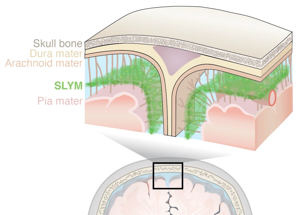
So, while dissecting a mouse for the study, Møllgård noticed something peculiar: a new body part. In the past, neuroscientists had established that there were three protective membranes between the brain and the skull. However, in this mouse, he spotted a fourth: a very thin membrane sitting just atop the brain. Later, they verified its existence in humans. The new membrane, called the subarachnoid lymphatic-like membrane (SLYM), is so delicate, it would be destroyed during most routine dissections—which is why it’s gone unnoticed until now.
The new membrane, called the subarachnoid lymphatic-like membrane (SLYM), works as a protective barrier and a filter for waste disposal.
University of Copenhagen
Nedergaard believes that the SLYM is an important structure that works as a protective barrier and a filter for waste disposal. All the brain’s waste is carried away by the cerebrospinal fluid (CSF), a watery substance that bathes the brain and spinal cord. Here the membrane plays an essential role.
“I think it [the SLYM] has a barrier function that separates clean and dirty CSF,” Nedergaard said. It may help collect, sort, and flush waste from brain cells—including debris, toxins, misfolded proteins, and byproducts of metabolism—separating waste and toxins from nutrients that can be reused or recycled.
The SLYM is also home to plenty of immune cells that can patrol and defend the brain, responding to infection or damage. Nedergaard posits that if there is a problem with the membrane, it could lead to further brain damage and neurodegenerative conditions like Alzheimer’s.
With scientists dissecting and studying brains every single day, why has it taken so long to spot the SLYM? The reason has to do with the fact that any fluid filled-spaces within the body collapse soon after we die. “Five minutes after we die, there’s almost no CSF left,” Nedergaard said. The brain absorbs it, expands, and ruptures any delicate structures in the way.
Luckily, Møllgård was doing a careful dissection of the mouse brain as he was searching for another type of delicate structure. Nedergaard’s lab had developed a mouse model to study a series of tunnels surrounding the brain called lymphatic vessels which are lined by immune cells called lymphocytes. These vessels are involved in transporting waste and other molecules out of the brain.
In their mouse model, the lymphocytes were marked with a fluorescent green protein which would make these tunnels visible after careful dissection. When the team looked, though, they discovered a layer of fluorescing green cells forming a thin layer on the top of the brain.
“I believe there is a great deal to discover about the microanatomy of the human body,” Paul Neumann, professor of medical neuroscience at Dalhousie University in Nova Scotia, Canada who was not involved in this study, told The Daily Beast. Finding these small structures may become more and more common as we continue to develop better microscopes and better techniques to study the human body.
“A function makes it, in my mind, super exciting. Something that can lead us to revise a textbook.”
— Paul Neumann, Dalhousie University
“We can now do in vivo microscopy,” Nedergaard said. This technique allows scientists to image humans and other animals while they’re alive, and visualize a group of cells—sometimes even watching them in action—by adding genes into the animals that tag a group of cells with fluorescent proteins. New discoveries like this get scientists excited about what the structures might mean for our understanding of the body.
“A function makes it, in my mind, super exciting. Something that can lead us to revise a textbook,” Nedergaard said.
Now her team is looking to understand the consequences of SLYM breakages as a result of disease or or traumatic brain injury. “I’ve no doubt that you would discover many more secrets about the body because not only are the techniques better, but we’re also asking more sophisticated questions,” she said.
In fact, we may be experiencing an anatomical renaissance where new, important structures are unintentionally discovered or rediscovered every few years.
Five years ago, surgeons were using an endoscope—a kind of snake-like camera—to look for cancer inside a patient’s bile duct. Instead of cancer, they found a series of pockets that no one had reported seeing before. After taking a closer look, they realized that these were collapsed structures—connected pockets below the skin and in the gut, lung, and urinary systems—and called it the interstitium.
“That was actually a major discovery,” Nedergaard recalled. “All the fluid is taken up by cells that swell up and that means that all fluid filled spaces disappear.” It turns out that the interstitium is important for regulating the immune system, and may be an important way that cancer cells spread through the body.
Sometimes what appears to be a new structure or organ in the body has been characterized decades or even centuries before, albeit with less sophisticated careful techniques. “One of my colleagues likes to say, ‘If you want to make a discovery, read an old book,’” Neumann said. In 1508, Leonard Da Vinci first characterized the mesentery, what appeared to be small membranes that helped the small intestine stick to the abdomen—think separate pieces of duct tape holding the gut in place.
For most of medical history anatomists believed that the mesentery was just another part of the gut because it wasn’t one continuous structure. But after careful examination in 2016, surgeons Calvin Coffey and Peter O’Leary from the University of Limerick discovered that the mesentery was one continuous membrane, a structure separate from the intestines. Treating it as its own organ opened up new insights into chronic gut disorders. For example, researchers discovered that cutting out the mesentery alongside diseased parts of the gut increases remission rates for Crohn’s disease.
Still, sometimes these structures are properly identified but disregarded by other scientists in the field. In 2012, the glymphatic system—large tunnels surrounding the brain’s blood vessels that carry waste molecules toward the lymph nodes—gained recognition as an important part of the brain’s immune system. However, it was actually discovered decades before.
“The discovery a decade ago of the ‘glymphatic system’ provided an explanation for observations reported by Helen Cserr in the late 1970s of clearance of material from the brain into lymph nodes,” Neumann said.
Cserr was a young researcher who wasn’t taken seriously, in part because no one thought the immune system affected the brain. “She produced a beautiful set of data in the 1980s showing the glymphatic system.” Nedergaard said. But other researchers were skeptical, and couldn’t replicate her work. “This big prominent lab published a couple of papers saying that she was wrong. She actually had to leave research.”
Cserr died in 1994 due to a brain tumor. It took three decades, and paradigm shifts in neuroscience—that the brain isn’t connected to the rest of the immune system— until her findings were proven right.
Møllgård had the past expertise to spot the new membrane. But in many cases, young researchers might discover something unexpected that ends up ultimately discarded. Nedergaard encourages her trainees to take a closer look at these unexpected findings because they are a learning opportunity, even when they turn out to be an error.
“And you also in some cases, when you can’t explain the finding, you keep it in the back of your mind,” she said. “Then maybe, it’s always a mystery, and maybe 10 years later, you have another observation, and you can put it together.”
The influx of advanced molecular biology, microscopic techniques, and inquiry will help us understand the minutiae within our bodies. This means that scientists will continue to accidentally discover, rediscover, and revise what we know about anatomy. Along the way, this may lead us to a few new avenues for better treatments for diseases alongside even more questions about our bodies.